Reuben Notes
Calcaneal Fractures
Majority of calcaneal fractures are a result of a fall from a height.
Most are intra-articular, which are more serious and disabling than intra-articular.
The area most commonly involved in calcaneal fractures is the posterior facet.
Anatomy
Diagnosis
Presentation & Mechanism
Classification
Imaging
Treatment Options
Surgery
Anatomy
Can be divided into 6 surfaces: supererior, inferior, medial, lateral, anterior, and posterior.
- Superior - Beak of calcaneus supports anterior facet, sustentaculum tali supports middle facet
- Lateral - flat with groove and trochlea centrally for the peroneal tendons. Impingement of the peroneal tendons are a common complication of lateral displacement (lateral wall "blow-out") of calcaneal fractures.
The calcaneus is composed of a shell of thin cortical bone. The inner cancellous bone displays a pattern which resembles the static and dynamic stresses placed on the bone.
- Traction trebeculae radiates from inferior cortex
- Compression trebeculae converge to support anterior and posterior facets
- Intersection of trebecular bone known as "thalamic portion of calcaneus".
-
Neutral triangle or Ward's triangle - area of sparse trebecular bone between compression and traction trebeculae.
- Lies beneath the angle of Gissane's and carries blood vessels to medullary cavity of calcaneus. This is the weakest portion of the calcneus as it is almost completely void of bone.
Superiorly:
- interosseous talocalcaneal ligament plays an important role in calcaneal fracture patterns.
Lateral
- Thin cortical wall (involved in blow outs)
- Soft tissue: can get tendonitis and nerve entrapment with fracture
Medially
-
Sustentaculum tali – keystone of calcaneal fracture repair
- Sustentaculum tali fragment – a.k.a. – anterior medial fragment, McReynold’s fragment, thalamic fragment
- Sustentaculum tali retains its position in fxs (maintains its anatomic relationship)
- Strong ligamentous attachment – deltoid, talocalcaneal ligaments and flexor hallucis longus à all play a role in maintaining the fx fragments
Anteriorly
- CC- joint – often have fractures that extend through the CC joint
Talar relationship to calcaneus
- Calcaneus is laterally displaced on the main body of the talus
- Force of ground pushes calcaneus up, most of weight is centered medially over the sustentaculum tali
Diagnosis
- Mondor's Sign - Hematoma or bruising which extends from the heel to the arch of the foot
- Injured side often wider than unaffected side - heel can appear flatter, wider and in a varus/valgus postition when compared to contralateral side
- Tenderness on both medial and lateral calcaneus with extreme pain on ROM of STJ
- Ankle joint ROM generally not painful
- Hoffa sign - loss of plantarflexion strength of the tendo achilles in severely comminuted fractures.
- Most cannot bear weight on heel
- Children may present with knee flexion and ankle equinus to avoid weight bearing
- Typically edema develops quickly after injury
- 10% of calcaneal fractures have compartment syndrome present - Manoli and Myerson
- Fracture blisters may be present
- Always evaluated other areas, particularly the lumbar and cervical spine, head, wrists, knees, and hips, for potential injuries
- Neurovascular - always evaluate neurovascular status, especially along course of sural nerve and posterior tibial artery
- Radiographs - DP of foot, Lateral of foot and ankle, Calcaneal axial. It may be difficult to appreciate the fracture as the architecture of calcaneus may mask the appearance of fractures.
-
-
Lateral
- best to assess Böhler's angle and crucial angle of Gissane
-
Böhler's angle - typically 25-40° - angle formed by intersection of lines from highest point of anterior process to highest point of posterior articular surface, and other line from same point on posterior articular surface to the most superior point of calcaneal tuberosity.
- Angle decreases or even reversed with severe fractures
- Crucial angle of Gissane - between 125-140° - created by subchondral bone of posterior facet and subchondral bone of middle and anterior facet
- Angle increased to greater than 180° with displacement of posterior facet in joint depression fractures
-
Böhler's angle - typically 25-40° - angle formed by intersection of lines from highest point of anterior process to highest point of posterior articular surface, and other line from same point on posterior articular surface to the most superior point of calcaneal tuberosity.
- The goal of adequate reduction are the recreation of both angles and restoration of the subtalar joint
- Medial oblique - used to visualize the involvement of the calcaneocuboid joint
- Broden I and II and the Isherwood II - used to evaluate the subtalar joint, especially the posterior facet - rarely used due to availability of CT scan
-
Lateral
- best to assess Böhler's angle and crucial angle of Gissane
-
- Lumbar spine - essential for patients who suffered a fall or patients who complain of back pain and have tenderness on physical exam of spine.
- CT scan - vital for evaluating extent of articular involvement
Presentation
Mechanism of Injury
-
Most common cause is
fall from a height
– often concomitant injuries – lumbar injuries or fractures
- Should get leg, ankle, and lumbar films to rule out concomitant injury
- Calcaneal fracture rarely occurs alone with a fall from a height
- MVA - Second most common
Plantar ecchymosis – hallmark of calcaneal fracture
- Not seen often with ankle fractures
- Calcaneus is vascular and bleeds quite a bit with a fracture
Marked edema
- Fracture blisters may occur
- Longer you wait to repair the increased incidence of infection and wound dehiscence due to edema
- Consider compartment syndrome – most compartment syndromes in feet are from calcaneal fractures
Pain around heel
Equinus gait
Mechanism of Fracture
Palmer – JBJS 1948
- Primary fracture line based on talus coming down on sustentaculum tali – shearing off of middle facet of sustentaculum tali
-
Primary fracture line
- Begins medially and exits superiorly through the posterior facet
- With continued compressive forces, the lateral process of talus impacts Gissanes angle
-
Secondary fracture line
- Continued forces push the posterior facet into body of calcaneus
- More vertical force – tongue fracture
- Most posterior force – same as Essex-Lopresti – joint depression fracture
Peter Essex-Lopresti – BJS 1952
-
Primary
fracture line
- Caused when there is a vertical force (from the ground up)
- Primary fracture line caused by the lateral process of talus impacting Gissane’s angle acting as a wedge to split calcaneus in half
-
Secondary
fracture line
- Occurs with continued compressive forces and body of talus is pressed into calcaneus – 2 secondary fractures can occur
-
Forces in
vertical nature
- Fracture exits calcaneus posteriorly – tongue fracture
-
More
posterior directed force
- Fracture line exits back up superiorly into the posterior facet – joint depression fracture
Resultant Deformity
- Joint incongruity – usually posterior facet
-
- Can lead to DJD
-
Decreased calcaneal height
compression of calcaneus
- limb length difference
-
Increased calcaneal width
- Soft tissue impingement – peroneal tendons and sural nerveà peroneal synovitis and sural nerve entrapment
- Can’t fit in shoe – too wide
Extra articular – better prognosis
Intra articular – worse prognosis
- >75% of all calcaneal fractures
Rowe
- Mainly for extra-articular fractures
-
Type I
-
A -
Fracture of the tuberosity of the calcaneus
- Best visualized on axial views
-
Always secondary to shearing forces
- Calcaneus is at an angle and forces shear off the tuberosity
- May be due to avulsion of plantar structures – fascia, intrinsic muscles
- If small and not grossly displaced do not need to be fixed
- If large or grossly displaced they need fixation
-
B -
Sustentaculum Tali Fracture
- Best seen on axial view
- Hallmark – pain with motion of FHL tendon, right under sustentaculum tali
- Shear type fractures
- May be avulsion from eversion sprain – deltoid ligament pulls of sustentaculum
- Don’t need to be fixed if in good approximation
- If grossly displaced they need fixation
-
C -
Fracture of the Anterior Process of the Calcaneus
-
2 main causes
- Avulsion from bifurcate ligament or EDB
- Compaction when there is dorsiflexion and inversion of the foot and the lateral talar process impacts the anterior process into cuboid
-
2 main causes
-
A -
Fracture of the tuberosity of the calcaneus
-
-
- If intra-articular and large they should be fixated for early motion and reduced chance of DJD at CC joint
-
-
Type II
- Fracture of posterior superior aspect of calcaneus
-
A -
Beak Fracture
- Dorsal 1/3 of tuberosity
- Shear or blunt trauma
- Lateral x-ray
- Don’t fix if small and non-displaced, just cast
- If large or displaced it needs fixation
-
B -
Avulsion Fracture at insertion of the achilles tendon
- Achilles avulsion – middle 1/3 of calcaneal tuberosity
- Often caused by dorsiflexion forces of foot
- Usually need fixation
- AK cast if well approximated
- If long standing (>3 weeks) they may need TAL or gastroc recession to get fragment down
-
A -
Beak Fracture
-
Type III
- Oblique Fracture of the Calcaneal Body NOt Involving the STJ
- Cast if not grossly displaced, if grossly displaced then it needs fixation
- Usually heal well due to vascularity of calcaneus
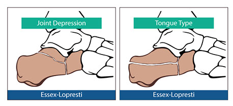
- Type IV - Intra-Articular Fractures Involving the Subtalar Joint (tongue-type - Essex-Lopresti)
-
Type V
- Intra
-Articular Central Depression with Varying Degrees of Comminution (joint depression - Essex-Lopresti)
- Depression of posterior facet into main body of calcaneus
- Severely comminuted
Essex-Lopresti - X-ray based classification
- Used for intra-articular
-
Tongue Type
- non- Joint depression
- Like Rowe Type IV
-
Joint Depression
- Like Rowe Type V
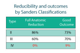
Sander's - CT classification (coronal)
- Number of fracture lines
- BEST - gives an idea of the type of clinical outcome and the ease of reducibility
-
Type I
- all non-displaced articular fractures, regardless of the number of fracture lines present
- No fractures through the posterior facet
- Type II - 2 part articular fracture of posterior facet - 1 fracture line
- Type III - 3 part articular fracture - Two fracture lines
- Type IV - 4 part articular fracture - Three fracture line
- Further classified into location of fracture lines
-
- A - Lateral fracture line
- B - Central fracture line
- C - Medial fracture line - Sustentaculum Tali Fracture
-
Reduction Criteria
for Sander’s Fractures:
-
Anatomic Reduction
- No incongruity in joint surface of posterior facet
-
Near
Anatomic Reduction
- <3 mm incongruity of joint surface of posterior facet
- An acceptable level of reduction
-
Approximate
Anatomic Reduction
- 3-5 mm incongruity of joint surface of posterior facet
-
Failed
Anatomic Reduction
- >5 mm incongruity of joint surface of posterior facet
-
Anatomic Reduction
Radiographs
Lateral Radiograph - Most important
-
Bohler’s Angle
-
20-45 degrees
- 1st line from most superior portion of posterior aspect of calcaneus to most superior portion of posterior facet
- 2nd line from most superior portion of posterior facet to anterior process
- Not indicative of severity, only indicated that there has been a fracture
- Get bilateral films to compare contralateral value
- May be subtle fracture, Bohler’s angle lets you know that there has been a change
- Look for flattening out of this angle
-
Gissane’s Angle
-
120-145 degrees
- Get flattening with fracture
- Angle measuring posterior facet and anterior facet.
Axial Radiographs - 2nd Most important x-ray
- Posterior aspect of tuberosity
- Lateral wall blowout
-
Relationship of sustentaculum tali to rest of calcaneus and calcaneal tuberosity
- Often get varus position of tuberosity
- Main body of talus is displaced medially which results in buckling
DP View (AP view)
- Anterior aspect of calcaneal fx
- Look at CC joint involvement
- Look for concomitant fractures elsewhere in the foot
Broden views
- Good for STJ congruity
- Looking at posterior facet
- Can give an idea of incongruity at posterior facet
- Patient lying supine on table
- Foot internally rotated 45 degrees
- X-ray beam tube head angles 10, 20, 30, and 40 degrees from perpendicular
- Helps you look at entire joint
CT Scan
- Best way to see Calcaneal fracture – A must for all Calc fractures.
- Gives 3-D view of what is going on in the calcaneus to know where all of the fragments are
-
Coronal view is most important on CT scan; sagittal and lateral
- Need to have knee flexed 50 degrees, foot 30 degrees, angle gantry at 15 degrees from perpendicular to cephalad to go straight through the foot
Treatment Options
- Cast without reduction – doesn’t address joint incongruity or deformity in shape of calcaneus
-
Cast with closed reduction
-
Bohler – took pin and placed medial to lateral through tibia
- Took another steinman pin an put medial to lateral through calcaneus
- Distracted to help reduce fracture to regain height, pulls calcaneus out of varus
- Used Bohler’s clamp – vice to help decrease width of calcaneus, applied medial to lateral to compress calcaneus
- Can cause soft tissue damage (nerve entrapments)
-
Gissane - more common
- Gissane spike – similar to steinman pin
- Placed spike from posterior to anterior through tuberosity and into the posterior facet fragment
- Pt prone with knee bent at 90 degrees then lifted leg off table to reduce fracture
- Forces posterior facet back up against talus and realigns calcaneus
- Casts around pin to maintain alignment
-
Bohler – took pin and placed medial to lateral through tibia
- Slipper cast modification – maintain STJ ROM for better healing – Essex-Lopresti modification
-
Primary arthrodesis – usually triple according to literature
- Only fuse affected joint unless several joints involved
- May be able to just STJ fusion
- If CC joint and posterior facet involvement – then do triple
- If pt has multiple comminutions then may want to primarily arthrodesis
- ORIF – treatment of choice for displaced intra-articular fracture
-
Techniques for repairing fractures are basically hybrids of 3 techniques:
- Essex Lopresti technique
- McReynolds technique
- Palmer technique
Open vs Closed Indications
-
Age
- chronological age may have no bearing on calcaneal fracture repair
- physiological age, activity level, goals and expectations are more important determinants
- osteoporosis is not a contraindication for fx fixation
-
Health status
- is patient surgical candidate, can they be NWB for 2-4 months, can they go through required rehab
- average intra-articular calcaneal fracture requires 1-2 years to get back to normal baseline activity
-
Activity level
- what was the patient activity level before the injury
-
Occupation
- take into consideration pts who work on there feet a lot
- prolonged recovery, but good outcome
- average intra-articular calcaneal fracture requires 1-2 years to get back to normal baseline activity
-
Intra-articular
- is there articular damage
-
Comminution
- fusion usually required if there is severe comminution
-
Good Clinical Outcome with ORIF
- Sangers – 73/70%
- Laughin – 78%
- Burdeax – 80.3%
- Thordarson – 80%
- Crosby – 47% - just did closed reduction
Goal of Reconstruction
- articular congruity reestablishment
- restore height and width of calcaneus
- allow early ROM otherwise there will be early increased risk of DJD, ankylosis, and stiffness
Fixation Methods
ORIF Essix-Lopresti
-
<50 years
- open if >50 then don’t ORIF due to healing potential
- correlated poor result with poor reduction – realized need for ORIF to properly reduce the posterior facet
-
advocated early ROM
- slipper cast
- related to successful outcome
-
Gissane spike – steinman pin – levers posterior facet back up against talus then applies cast to hold in place
-
if
tongue fracture
– pin percutaneously, closed procedure
- posteriorly into tongue then lever up the fx fragments
-
if
joint depression
fracture – did through open lateral incision but same reduction technique
- could visualize placing spike into posterior facet
- still commonly used
-
if
tongue fracture
– pin percutaneously, closed procedure
ORIF – McReynolds
- medial approach to fixate calcaneus
-
importance of sustentacular fragment (McReynold's Fragment)
- 1st to realize importance of sustentaculum fragment
- once compressive force from injury is released the sustentaculum and talus recoils because of deltoid ligaments and interosseous attachments of the sustentaculum tali to talusà keep calcaneus in position
- sustentacular fragment remains anatomic and is cornerstone for repair
- put the rest of the pieces back based on where the sustentacular fragment is located
-
negatives
- a portion of the facet may stay with sustentaculum while the rest in shoved down into the main body of the calcaneus – step off fracture
- more invasive approach
- must watch for medial neurovascular structures and tendons
- must go through medial wall of calcaneus
- not as commonly used
- remember to always build the calcaneus around the fx
ORIF – Palmer
- lateral approach
- raise posterior facet back up to anatomic position
- bone graft to buttress posterior facet after repositioning facet
- most commonly used today
Surgical Considerations
-
Edema
- need to do within hours (1st 8-12 hrs) of fracture (before there is gross edema) or wait (5-10 days) until edema has resolved
- edema will cause dehiscence or difficulty in closure and causes tension on soft tissue
- lateral incisions often dehisce anyway
- if edema is already present it must be brought down before fracture is repaired
- elevation
- ice: won’t bring the current edema down, but future edema is managed
-
compression
- compression pump to actively pump edema out of foot
-
Fracture blister
- can operate right through fracture blister
- result of gross edema
-
Antibiotic prophylaxis
- hematoma and lots of dead space from compaction
- usually long procedure – 2 hours
- putting in lots of hardware
- can have high incidence of infection so must prophylax
- 1g ancef pre op- and 1g q8h for 24 hours post-op
-
usually kept for 2-3 days in the hospital
- hemostasis, edema control, and antibiotic prophylaxis
-
Incision planning
- must plan incision around additional wounds or fractures
- lateral skin is tenuous more likely to dehisce
-
Bone graft use
- if there isn’t sufficient bone stock for fixation a bone graft may be needed to hold the posterior facet in place
- defects can fill in quickly on their own due to the vascularity of the calcaneus
- depends on size of defect; do you need additional support
Surgical Technique
- General/spinal anesthesia – paralysis of lower extremity for easier reduction, long case
- Thigh tourniquet
-
Usually lateral incision – lateral extensile incision (“L” shaped incision)
- L incision – l incision beginning just posterior to lateral mallelolus extending to base of 4th and5th met base area
- L incision creates flap for greater exposure (move as one big flap)
- incision parallels peroneal tendons and sural nerve, keep incision posterior and inferior to peroneal tendons and sural nerve to keep these structures within the flap
- make incision all the way to bone (no layering) à decreases incidence of dehiscence, the more layers there are the greater the incidence of dehiscence
- lateral wall blow out gives natural window into calcaneus
- atraumatic technique
- Use no-touch retraction – suture flap open or use k-wires, don’t keep pulling on flap
- Reflect pieces of lateral wall that blew out to get to posterior facet
- Use freer elevator to lever the posterior facet back to its position
- Use bone curette and remove all of hematoma, can also use pulsed lavage
-
Reduce other fractures – tuberosity (get out of varus position using spike))
- tuberosity – may need k-wire, recreate fracture, distract and reduce fracture
-
When fixating use fluoroscopy – if fixation it too far it may go into tendons or neurovascular bundle
- temporarily fixate with K-wires through posterior facet into sustentaculum tali
- Place joint into ROM to look for incongruity
-
Permanently fixate – calcaneal reduction plate (Sander’s Plate), several different plates can be used, just reduce the fracture – 1st screw is usually the one through the posterior facet
- fixate into sustentacular fragment – from lateral to medial, holds articular surface in position with the STJ,
- if there is not enough bone stock, a bone graft of appropriate size can be used to buttress the posterior facet in position
- can use screws, k-wires, or bone staples to fixate
- Close in 2 layers – periosteum (if present); the skin flap
- Always use drain – closed suction drain
- Light compressive dressing
- Keep overnight – draining, hemostasis, neurovascular status, prophylactic antibiotic
-
Post-Op Care
-
early ROM – one consensus with in the literature
- begin when wound is beginning to heal to decrease chance of dehiscence – 10 days
- use removable cast boot to start early ROM
-
delayed weight bearing
- 3 months average depending on extent of fracture, comminution, and loss of bone stock
- 8-12 weeks, usually 12 weeks NWB
- follow radiographically
- WB for 1 month (4 weeks) in cast – CAM walker – when there are signs of osseous healing
- WB in shoe
- usually 3-4 months to get in shoe but still have disability and pain
- may take 1-2 years for full rehabilitation
-
early ROM – one consensus with in the literature
Complications
- heal
-
infection
- flush periodically during surgery, remove hematoma, antibiotic prophylaxis, good atraumatic technique, need to consider especially if patient is immunocompromised
-
Joint stiffness
- biggest complication, should be expected with calcaneal fracture, nature of the fracture
-
DJD
- Intra-articular fracture – will occur, may eventually need STJ fusion, if calcaneus has been repaired it will be easier to later fuse
- importance of congruity and early ROM
-
soft tissue impingement
-
peroneal tendons and sural nerve from lateral wall blow-out and widening of calcaneus
- peroneal tendon synovitis and sural nerve entrapment
-
peroneal tendons and sural nerve from lateral wall blow-out and widening of calcaneus
-
mal-position
- repair technique, reduce varus position of tuberosity
- also be aware of height and width
- a fluoroscopy a must
-
non-union
- rare due to vascularity of calcaneus (he has never seen this)
-
heel pad damage
- usually due to disruption of fibrous septae from impact from fall
-
RSD
- depends on extent of trauma
-
compensatory injury
- pain and stiffness will lead to altered gait which may cause injury elsewhere
-
compartment syndrome
- common